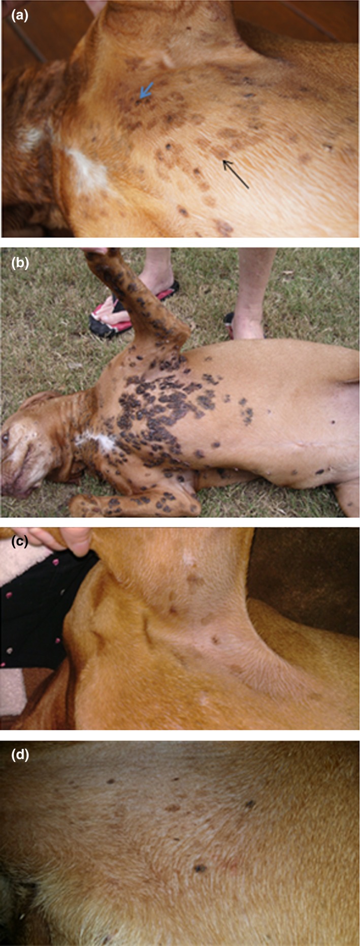
Figure 1.

Appearance of pigmented papillomas on Case 1, 2 & 3. (a) shows lesions in Case 1 in 2011, while the lesions observed in 2015 are illustrated in (b). Comparing (a & b) provides an appreciation of the slow but insidious progression of lesions over time. In (a), small pigmented plaques (3–8 mm diameter) on the skin of the sternum and ventral cervical region are highlighted by a narrow black arrow in one instance; approximately 10% of the lesions appeared exophytic (e.g. blue arrow), consisting of dark grey pigmented proliferations. (c) shows representative lesions from Case 2 in 2014, while (d) illustrates lesions from Case 3. Note Case 1 (a & b) is much more severely affected than Cases 2 (c) & 3 (d).
