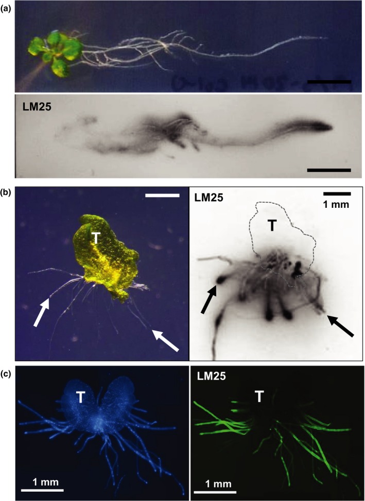
Figure 2.

Detection of xyloglucan (XG) secretion from plant surfaces with XG MAb LM25. (a) Bright field image of Arabidopsis seedling grown on plant agar solid media for 14 d paired with nitrocellulose print of solid media surface (after removal of the seedling) which was then probed with LM25. Bars, 10 mm. (b) Secretion by day‐30 Marchantia polymorpha gemma. Bright field image on agar and immunoprint of agar surface after gemma removal. Arrows indicate corresponding rhizoid tips. T, thallus outlined in print by dashed line. Bars, 1 mm. (c) Whole mount immunolabelling of day‐14 M. polymorpha thallus/rhizoid in situ on agar block with LM25. Blue represents Calcofluor White labelling of cell walls and green represents LM25‐FITC. T, thallus. Bars, 1 mm.
