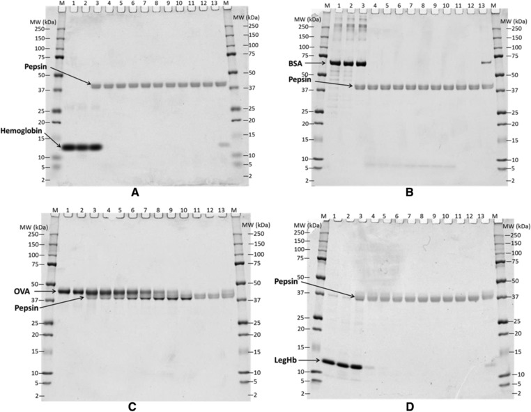
Figure 3.

Coomassie Brilliant Blue Stained SDS‐PAGE Gel Showing the Digestion of control samples, Bovine Hb (A), BSA (B) and OVA (C), and LegHb preparation (D) in SGF at the ratio of 10 Units pepsin per μg test protein (pH 2.0). All proteins were loaded 1.47 μg per lane as predigestion concentration. Lane M, molecular weight marker; Lane 1, protein control at 0 min; Lane 2, protein control at 60 min; Lane 3–10, digestion at 0, 0.5, 2, 5, 10, 20, 30, and 60 min; Lane 11, pepsin control at 0 min; Lane 12, pepsin control at 60 min; Lane 13, 10% of undigested protein.
