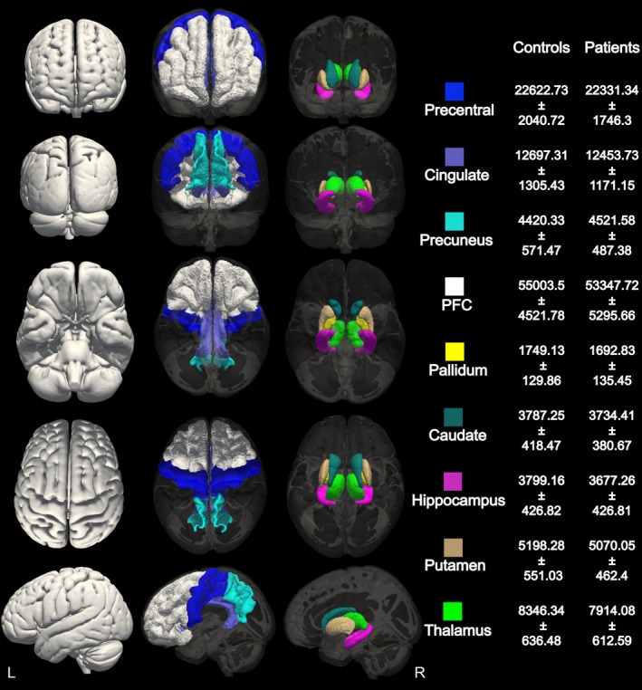
Figure 1.

Cortical and subcortical volumetric segmentation. Gray matter masks of the cortical and subcortical areas selected for volumetric analysis between patients and controls. Average volumes and standard deviations for each area in the healthy control and AED‐naive group are reported in mm3
