
Figure EV2. Monensin‐induced LC3 lipidation to entotic corpse vacuole requires the WD40 CTD of ATG16L1.
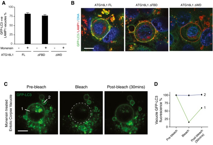
- Quantification of GFP‐LC3 recruitment to LAMP1‐positive entotic corpse‐containing vacuoles in MCF10A ATG16L1 −/− cells complemented full‐length (FL), ΔFBD or ΔWD ATG16L1 ± monensin (100 μM, 1 h).
- Representative confocal images of entotic corpse‐containing vacuoles in GFP‐expressing MCF10A cells treated with 100 μM monensin for 1 h and stained for LAMP1 (red) and DNA (blue). Scale bar: 10 μm.
- Representative sequence images from FRAP analysis of GFP‐LC3 on entotic corpse‐containing vacuoles treated with monensin (100 μM, 1 h). The region marked by a broken‐line circle was photobleached, and the recovery of fluorescence at line 1 and 2 was monitored. Scale bar: 10 μm.
- Quantification of GFP fluorescence at line 1 and 2 from (C).