
Figure 4. The WD domain of ATG16L1 is essential for LC3‐associated phagocytosis and other non‐canonical autophagy‐dependent processes.
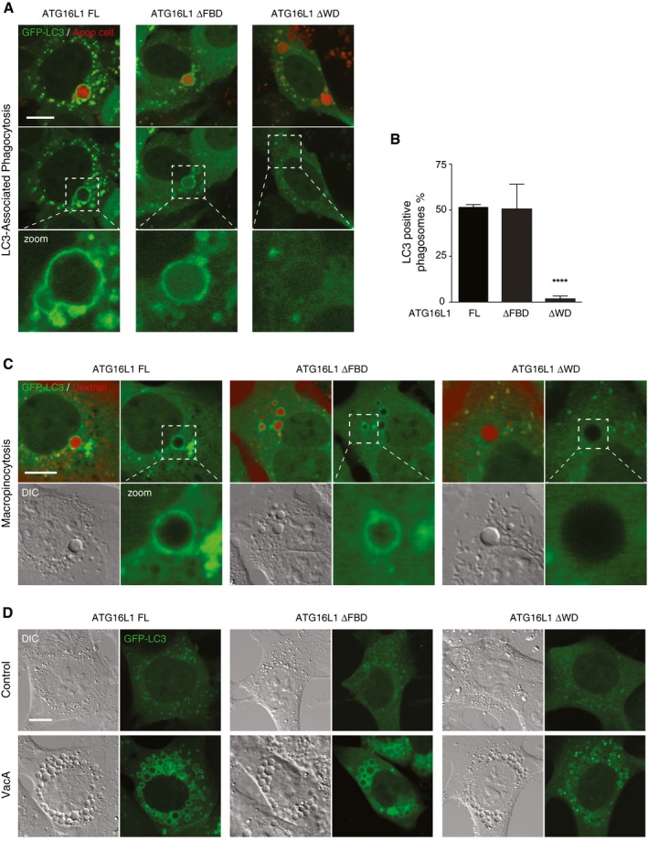
- Confocal images of GFP‐LC3 in ATG16L1‐complemented MEF cells phagocytosing red‐labelled apoptotic cells. Scale bar: 10 μm.
- Quantification of GFP‐LC3 recruitment to apoptotic corpse‐containing phagosomes in (A). Twenty phagosomes were counted per experiment.
- Confocal images of red dextran‐positive macropinosomes ATG16L1 complimented MEF cells. Cropped images show macropinosomes. Scale bar: 10 μm.
- Confocal images of GFP‐LC3 in ATG16L1‐complimented MEF cells treated with VacA toxin (10 μM, 4 h). Scale bar: 10 μm.