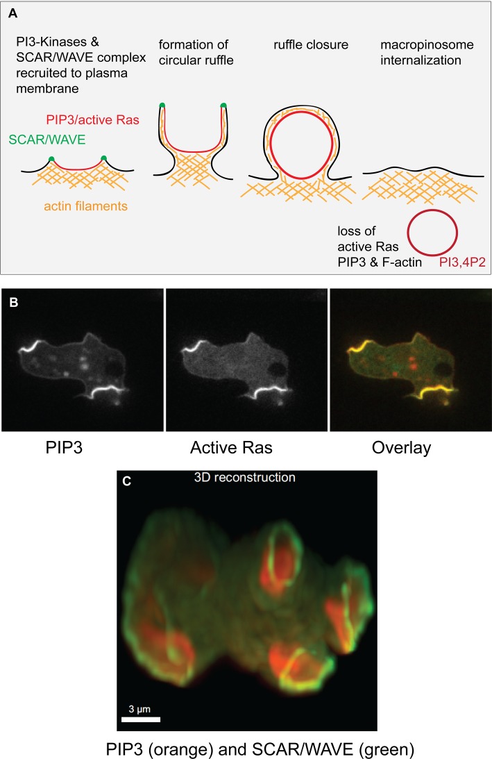
Figure 1. PIP3 and active Ras patches organize macropinocytic cups in the plasma membrane of Dictyostelium cells.
(A) Schematic diagram of macropinosome formation, showing the extension of a circular ruffle organized around a patch of intense PIP3 and active Ras in the plasma membrane. The circular ruffle can be several microns in diameter and eventually closes to create a macropinosome, which loses its PIP3, active Ras and F-actin. The PIP3 is converted into PI3,4P2 and the vesicle is trafficked into the cell. This diagram is based on Dictyostelium work; in mammalian cells, the importance of PIP3 for macropinosomes is well established, but that of Ras and SCAR/WAVE less so. It should be noted that Dictyostelium phosphoinositides are chemically unusual, being ether-linked, plasmanylinositols [28] but appear functionally equivalent to their mammalian counterparts. (B) PIP3 and active Ras form coincident, intense patches in the plasma membrane. (C) The SCAR/WAVE complex (green) is recruited to the periphery of these patches (reported by PIP3, red) where it activates the Arp2/3 complex to trigger actin polymerization. This provides the template for the walls of a macropinocytic cup. The images show growing Ax2 cells either in section or 3D-rendered, expressing reporters derived from PH-CRAC for PIP3, RBD of Raf1 for active Ras and HSPC300 for the SCAR/WAVE complex. Taken from ref. [21].

