Figure 4.
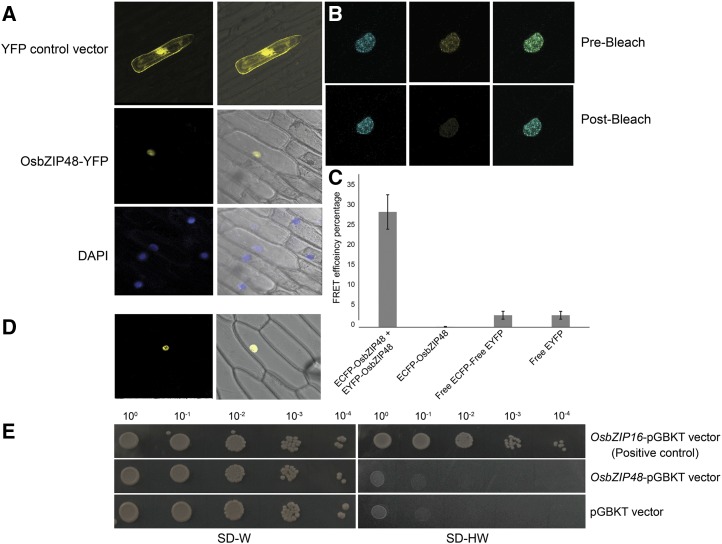
OsbZIP48 is localized in the nucleus, forms a homodimer, and lacks transactivation activity. A, Particle bombardment of the YFP-OsbZIP48 construct in onion cells. The first column shows photographs taken in dark field, and the second column shows merged photographs of dark field and bright field captured using a Leica microscope. The first row (YFP control vector) shows localization of only YFP protein, the second row (OsbZIP48-YFP) shows localization of OsbZIP48 tagged to YFP protein, and the third row (DAPI) shows DAPI-stained nucleus. B, Prebleach and postbleach images showing bleaching of YFP-OsbZIP48 for FRET analysis. C, Histogram showing FRET efficiency of CFP-OsbZIP48 and YFP-OsbZIP48 interaction as compared with the controls. Data shown are means ± se; n = 10. D, BiFC analysis using onion peel cells showing the homodimerization of nEYFPC1-OsbZIP48 and cEYFPC1-OsbZIP48 in the nucleus. E, Transactivation assay of OsbZIP48 in yeast cells. OsbZIP48 lacks transactivation activity, as the yeast cells containing the OsbZIP48-pGBKT construct were unable to grow on SD-HW medium (synthetic defined medium without histidine and tryptophan amino acids).

