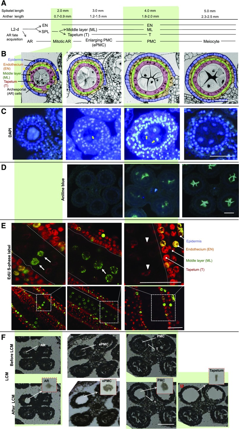
Figure 1.
Morphological and histochemical analysis of maize B73 anthers in 2- to 5-mm spikelets and the isolation of male germinal cells using laser microdissection. A, Developmental progression of L2-d cells in maize anthers in spikelets of given lengths. B, Cross sections of anther lobes at the stages analyzed. Yellow arrowheads point to the positions where only three concentric cell layers can be observed. AR, Archesporial cells; EN, endothecium (yellow); ePMC, enlarging pollen mother cells; ML, middle layer (green); PMC, pollen mother cells; SPL, secondary parietal layer; T, tapetum (pink). C, Cross sections of anthers stained with DAPI. Yellow arrows point to nucleolus. D, Cross sections of anthers stained with aniline blue. E, Longitudinal sections of anthers stained with EdU (green signal). Arrows indicate EdU-labeled S-phase cells. Arrowheads point to cells without EdU signal. F, Isolation of male germinal cells and tapetal cells by the Zeiss PALM microdissection system. Representative pictures of anther sections before and after laser capture were shown. Insets: Cells captured. Bars = 50 μm.

