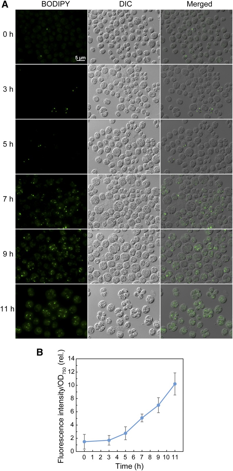
Figure 2.
Neutral lipid accumulation in Chlamydomonas CC1010 cells subjected to high-light irradiation. A, Observation of BODIPY-stained cells. The cells grown under low light were shifted to high light and grown for 11 h. DIC, Differential interference contrast microscope image; Merged, merged image of BODIPY and differential interference contrast microscopy images. B, Measurement of the fluorescent signal of Nile Red. Cells grown as in A were stained with Nile Red, and the fluorescent signal was measured by a plate reader.

