Figure 3.
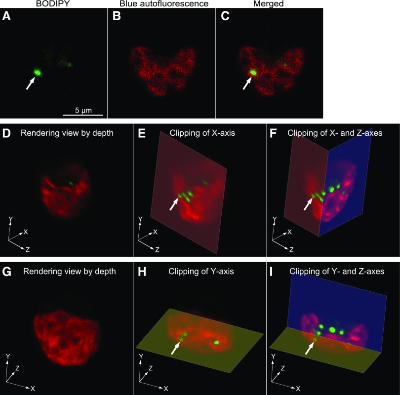
3D reconstruction by confocal fluorescence microscopy of a wild-type Chlamydomonas CC-1010 cell after the transfer from low light to high light. Cells grown under high light for 7 h were stained with BODIPY and observed by confocal microscopy with Z-stacking. The fluorescence of BODIPY and blue autofluorescence are pseudocolored. A to C, 2D images of a slice in which a lipid droplet (white arrow) seemed to be entirely enclosed by the chloroplast. Images A and B show the fluorescence of BODIPY and blue autofluorescence, respectively. Merged, Merged image of BODIPY and autofluorescence images. D to I, Rendering images of the same cell as the one shown in A to C. D to F show the images viewed from an identical angle. G to I show the images viewed from a different angle. E and F are clipped images of D at the x axis (E) or the x and z axes (F). H and I are clipped images of G at the y axis (H) or the y and z axes (I). The white arrows indicate an identical lipid droplet in A, C, E, F, H, and I.

