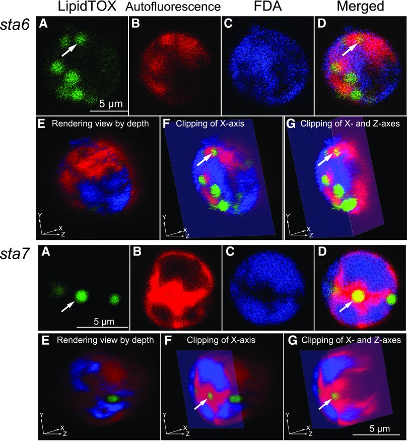
Figure 6.
3D reconstruction by confocal fluorescence microscopy of the starchless mutant cells of Chlamydomonas, deprived of nitrogen for 24 h with acetate supplementation. The top set of images shows a representative cw15sta6 cell; the bottom set of images shows a representative cw15sta7 cell. All images are pseudocolored. A, Localization of lipid droplets detected by LipidTOX fluorescence (green). B, Localization of chloroplast detected by blue autofluorescence (but shown in red). C, Localization of cytosol detected by FDA fluorescence (blue). D, Merged images of A to C. E to G, Views of 3D reconstructed images by different clipping. The staining with FDA in living cells will give uniform fluorescence after a long time due to hydrolysis of FDA by esterases leaked from broken cells. We had to work rapidly with the lowest concentration of FDA. That is why the intensity of fluorescence was weak in this observation.

