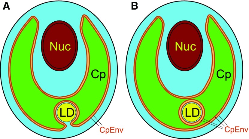
Figure 7.
Models of the localization of lipid droplets in Chlamydomonas. A, Plausible model of a lipid droplet seemingly located within a chloroplast but localized within the cytosolic compartment, which is present in a chloroplast invagination. The close association of the chloroplast envelope and the lipid droplet half-membrane supports the transfer of fatty acids from the chloroplast to the lipid droplet. B, Hypothetical model of a lipid droplet seemingly embedded within a chloroplast. The cytosol is shown by cyan. Note that the lipid droplet is surrounded by cytosol. Cp, Chloroplast; CpEnv, chloroplast envelope; LD, lipid droplet; Nuc, nucleus. This figure shows only a lipid droplet seemingly present within the chloroplast (type C localization). Besides this type of lipid droplet, there are a number of lipid droplets located in the cytosol between the nucleus and the chloroplast (type A) or outside the chloroplast (type B).

