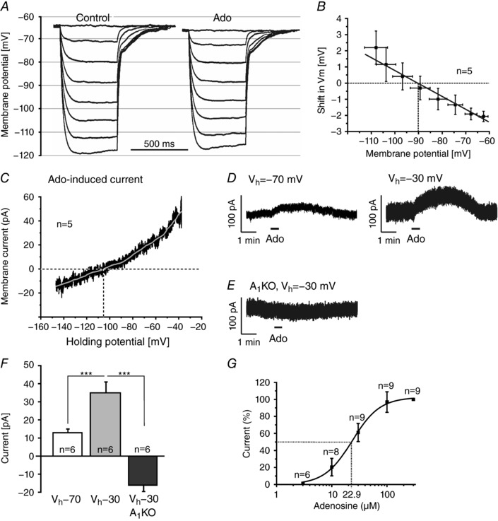
Figure 4. Adenosine activates potassium channels.

A, shifts in membrane potential evoked by negative current injections (−50 to −350 pA) before (control) and in the presence of adenosine (100 μm). B, extrapolation of adenosine‐evoked membrane potential shifts at different baseline potentials (as measured during the current steps in control) revealed an average reversal potential of −90 mV. C, current–voltage relationship of the adenosine‐evoked current, averaged over recordings from five cells (black trace) and filtered (grey trace; Savitsky‐Golay with second order polynomial and window size of 1501, corresponding to ± 5 mV), revealed a reversal potential of −106 mV. D, outward current evoked by 100 μm adenosine at a holding potential (V h) of −70 mV and −30 mV. E, adenosine failed to evoke outward currents in A1 receptor knockout mice. F, the adenosine‐evoked outward current was significantly larger at V h of −30 mV compared to −70 mV. At −30 mV, adenosine evoked an inward current in A1 knockout mice. G, dose–response relationship of the adenosine‐evoked outward current.
