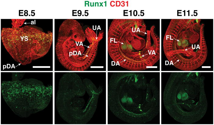
Figure 1. Location of Runx1 expression and hemogenic endothelium in the mouse embryo.
Confocal Z-projections of mouse embryos between embryonic day (E) 8.5 and E11.5 immunostained for the endothelial and hematopoietic marker CD31 (red) and Runx1 (green). Runx1 is expressed in hemogenic endothelium in the yolk sac (YS) at E8.5. At E9.5, Runx1 expression is prominent in the vitelline artery (VA) and umbilical artery (UA). An E10.5 embryo (head removed) shows Runx1 protein in the vitelline artery, umbilical artery, dorsal aorta (DA), and the site of colonization, the fetal liver (FL). al, allantois ; pDA, paired dorsal aorta. Scale bar = 500μm.

