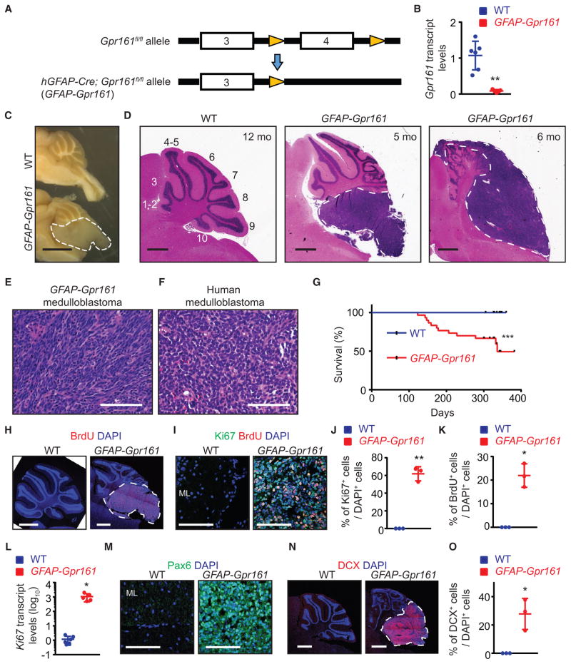
Figure 1. Gpr161 Deletion in Neural Stem Cells Induces Cerebellar Tumorigenesis.
(A) Schematic showing Gpr161 exon 4 deletion with the hGFAP-Cre allele.
(B) Gpr161 transcript levels in tumors of GFAP-Gpr161 cko (hGFAP-Cre; Gpr161fl/fl) mice (n = 5) are significantly decreased compared with the cerebellums of littermate controls designated as wild-type (WT) (Gpr161fl/fl or Gpr161fl/+; n = 6) collected from 3- to 5-month-old mice.
(C) Representative picture showing brains of GFAP-Gpr161 cko and WT mice (both 10 months old) after sectioning at the midline. Note the tumor (white dotted line) in the posterior cerebellum.
(D) H&E-stained sagittal sections showing tumors (white dotted lines) in the posterior cerebellum of GFAP-Gpr161 cko mice (5–6 months old).
(E and F) H&E-stained section of tumor from GFAP-Gpr161 cko mice (E) resembling that of human classic medulloblastoma (F).
(G) Kaplan-Meier survival curves of control (n = 19) and GFAP-Gpr161 cko mice (n = 27). Data were collected when mice were found dead or mice were sick (hunched back and reduced mobility).
(H and I) Representative images of immunostaining for BrdU (upon 1-h pulse) (H) and Ki67/BrdU (I) in the cerebellum of 3- to 5-month-old mice.
(J and K) Quantification of Ki67+ (J) or BrdU+ (K) cells co-stained for DAPI per field of view. n = 3 fields/mice, 3 mice/genotype.
(L) qRT-PCR analysis of Ki67 transcript levels in the GFAP-Gpr161 cko tumors (n = 5) with respect to normal cerebellum from the WT (n = 6).
(M) Pax6+ cells were present in the GFAP-Gpr161 cko tumor but not in the WT molecular layer (ML).
(N and O) Representative images of doublecortin (DCX) immunostaining (N) and percentage of DCX+ cells co-stained for DAPI per field of view (O) in the tumors of GFAP-Gpr161 cko mice and cerebellum of the WT. n = 3 fields/mice, 3 mice/genotype, 3–5 months old.
Data represent mean ± SD. *p < 0.05, **p < 0.01 by log rank test (G) and Student’s t test (B and J–L, and O). Scale bars indicate 2mm(C), 1mm (D, H, and N), and 100 μm (E, F, I, and M). (H) and (N) are tiled images. See also Figure S1.

