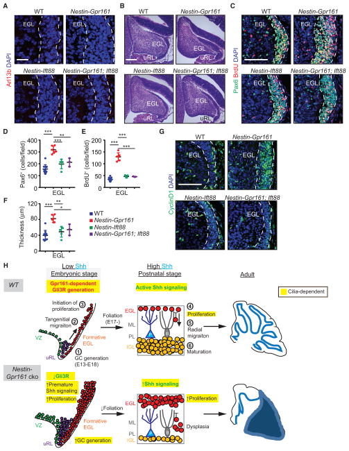
Figure 7. Gpr161 Prevents Premature Shh Signaling during Embryogenesis in a Cilium-Dependent Manner.
(A) Arl13b+ primary cilia in the formative EGL (white dashed line) at E15.5.
(B) H&E-stained sagittal sections of the embryonic cerebellum at E15.5.
(C–F) Representative images of proliferating GC progenitors in the formative EGL at E15.5 (C) and quantification (D and E). Also shown is the thicknesses of the EGL at E15.5 (F). The numbers of mice were as follows: WT (n = 11), Nestin-Gpr161 cko (n = 6), Nestin-Ift88 cko (n = 5), and Nestin-Gpr161;Ift88 dko (n = 3).
(G) Increased number of Cyclin D1+ cells in the formative EGL of Nestin-Gpr161 cko mice at E15.5.
(H) Cartoon summarizing the role of Gpr161 in cerebellar development and Shh-MB pathogenesis. See Discussion for details.
Scale bars indicate (A and B) 200 μmand (C and G) 100 μm. Nuclei are stained by DAPI. *p < 0.05, **p < 0.01, ***p < 0.001 by one-way ANOVA with Sidak multiple comparisons test. Abbreviations of genotypes are as in Figure 6. See also Figure S6.

