
Fig. 2.
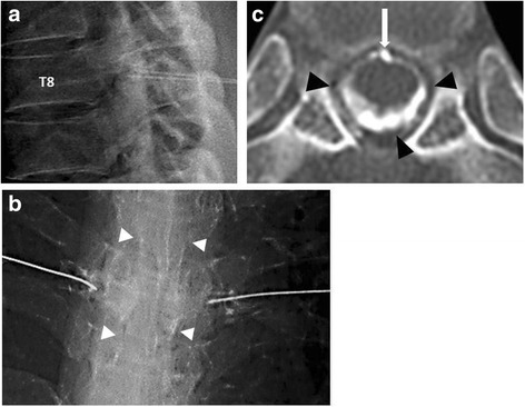
a Fluoroscopy-guided transforaminal EBP through bilateral T8 nerve roots (lateral view. b After contrast medium injection to confirm the epidural space (white arrowheads) (anteroposterior view. c Post-TFEBP spinal CT image at T7/T8 disc level demonstrating extension of injected blood through the entire epidural space (black arrowheads).The white arrow shows a partially calcified disc