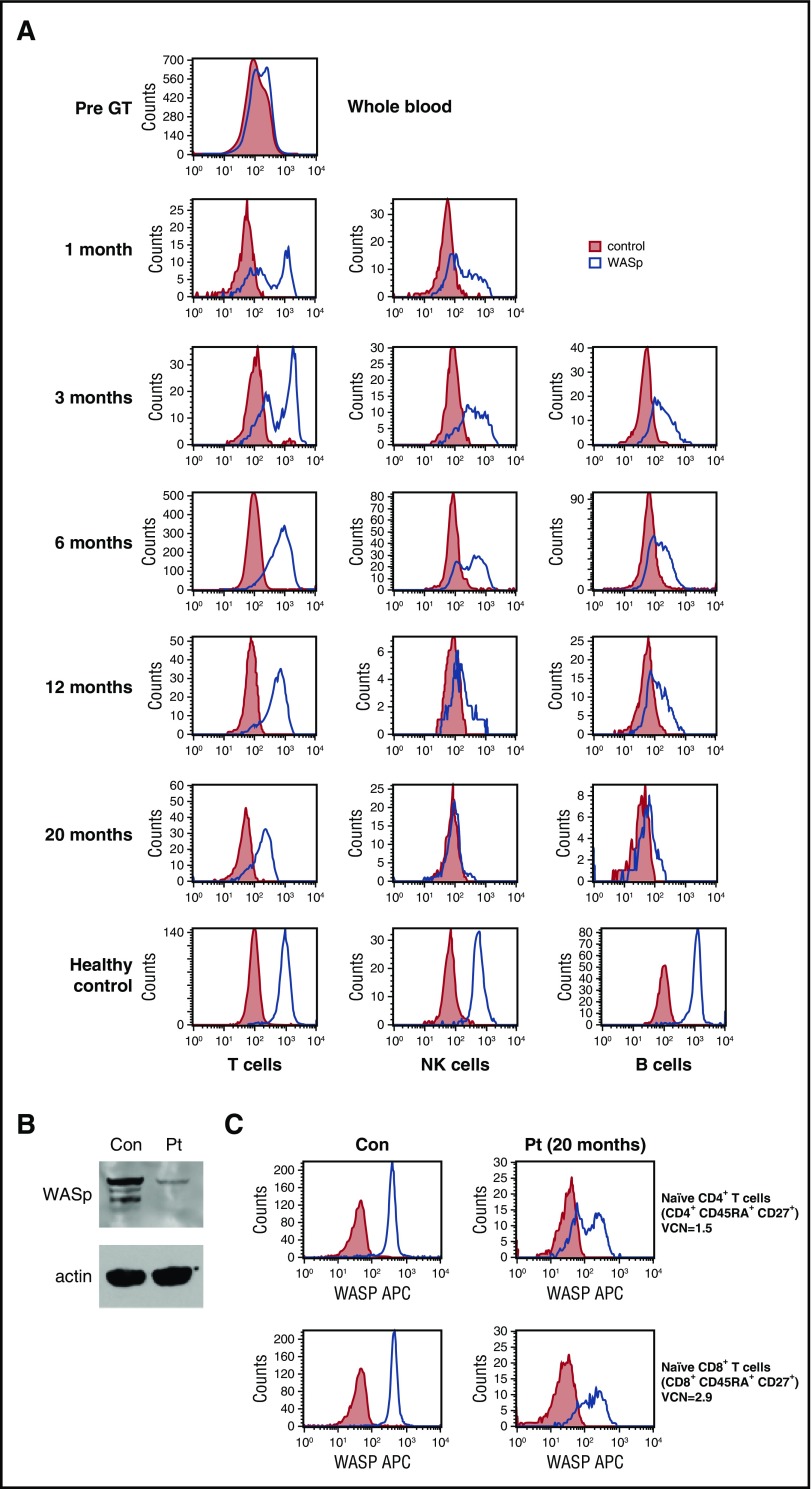
Figure 2.
Correction of WASp expression post-GT. (A) WASp protein expression in T cells, B cells, and natural killer cells. (B) Western blot demonstrating WASp expression in the patient’s platelets (Pt) compared with a healthy control (con). NB, patient received no platelet transfusions at any time. (C) WASp expression in purified naive CD4+ and CD8+ T cells at 20 months post-GT.

