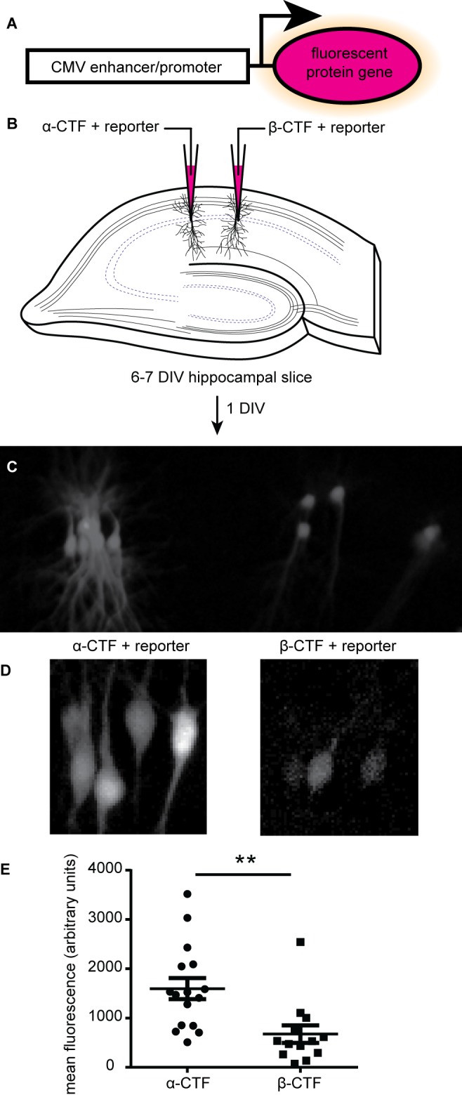
Figure 1. Detecting the effects of amyloid-β on CA1 hippocampal neurons using a CMV:GFP reporter.

A. Schematic of the reporter. B. Depiction of electroporation of hippocampal slices with α-CTF or β-CTF expression plasmids and the reporter plasmid. C. Epi-fluorescent microscope image of an electroporated hippocampal slice one day after electroporation. D. Two-photon microscope merged z-stack images from the same slice as in (C). E. Mean fluorescence from the fluorescent reporter protein (n = 16 for α-CTF, or 13 for β-CTF, mean ± SEM, **unpaired t-test P value < 0.01).
