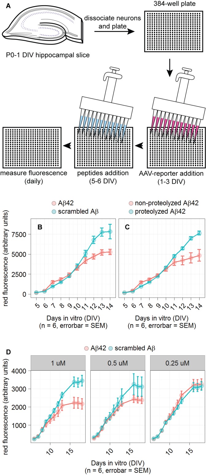
Figure 2. Detection of Aβ42 in primary neuronal cultures.

A. Schematic of the reporter system in 384-well plates. B and C. Plots of dsRed reporter fluorescence when cultures were incubated in the designated peptides. Primary neuronal cultures grown in 384-well plates were infected with reporter-containing AAV added at 1 DIV. Indicated peptides were added at 5 DIV, just prior to first plate reader measurement. D. Testing three concentrations of peptides: 1, 0.5, and 0.25 µM. NMDA was added to 200 µM at 11 DIV in an unsuccessful attempt to enhance reporter fluorescence. n indicates number of wells used for each condition and the error bars represent standard error of the mean (SEM). For all conditions using 1 µM peptide, the difference between reporter fluorescence for Aβ42 and scrambled Aβ conditions had an unpaired t-test P value < 0.05 on the last day of measurement.
