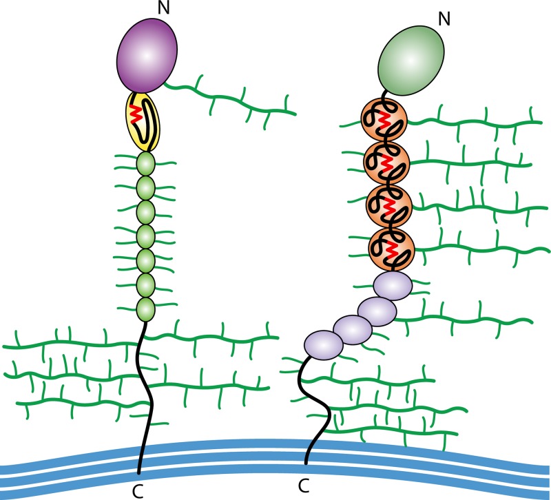
FIG 3.
Architecture of yeast adhesins. Shown are simplified cartoons of an Als adhesin from C. albicans (left) and Flo1p from S. cerevisiae (right). Each protein has a well-folded β-sheet N-terminal domain (NTD) (purple or green), amyloid-forming sequences (red), tandem repeats (repeated colored ovals), an unstructured stalk region, and a C-terminal cross-link to cell wall β-glucan (blue). N- and O-glycosylations are shown in green.

