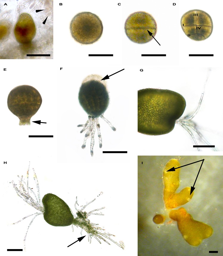
Fig 4. Early development of Cystoseira amentacea var. stricta.
A. Detail of a conceptacle with an oogonium and antheridia (arrowhead). B. Zygote with a central large nucleus. C. First zygote division (arrow). D. Second zygote division (II) parallel to the first (I) and third (III) and fourth divisions (IV) perpendicular to the first. E. Embryo with rhizoidal buds (arrow). F. Embryo with secondary rhizoids. Note the detachment of the fecundation membrane (arrow) during embryo elongation. G. Hyaline hairs growing from the invagination in the apical region of the embryo. H. Embryo with long apical hairs and numerous rhizoids (arrow). I. Germling with cryptostomata (arrows). Bar = 200 μm.

