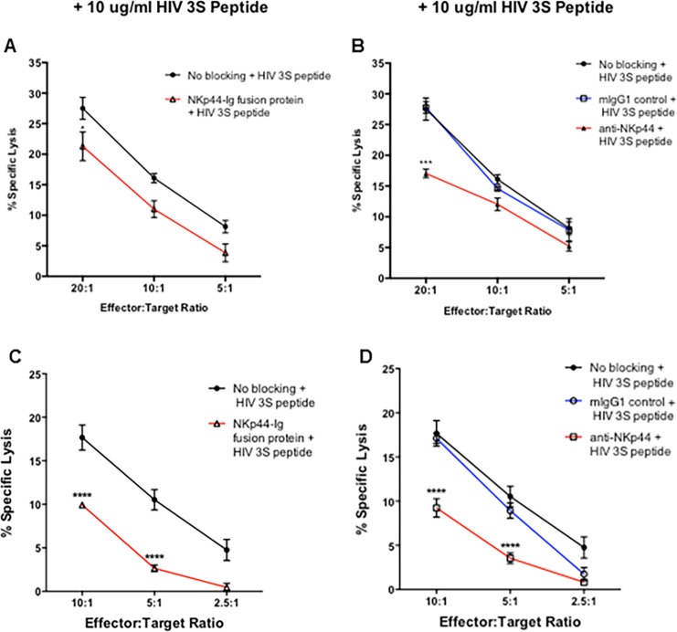
Fig 4. HIV-3S peptide stimulation protects astrocytes and the blocking of NKp44 further protects HIV-3S stimulated astrocytes from NK92-MI and primary NK cell mediated killing.
Astrocytes were stimulated with 10 μg/ml HIV 3S peptide for 4 hours prior being labeled with 51Cr. NK92-MI and primary NK cell mediated lysis of astrocytes was determined using a standard 51Cr release assay. Astrocytes and NK92-MI cells (A & B) / primary NK cells (C & D) were first blocked with Human IgG Fc fragment to prevent reverse binding of fusion protein and antibody dependent cellular cytotoxicity. Astrocytes cells were labeled with 51Cr and incubated with 1 μg/μl of NKp44-Ig fusion protein (A, C). Astrocytes were then incubated with NK92-MI and primary NK cells at varying effector to target cell ratios for 4 hours at 37°C. Percent specific lysis of astrocytes was compared to astrocytes incubated with 0.5 mg/ml mIgG1 isotype antibody or no antibody, which served as a positive control (No Blocking) of cell lysis under unblocked conditions. Alternatively, NK92-MI and primary NK cells were incubated with 0.5 mg/ml anti-NKp44 or mIgG1 isotype control antibody prior to incubation with astrocytes incubated with no antibody (B, D). Figure is representative of 3 independent experiments performed in triplicate. Data is displayed as means ± SEM. ** p < 0.01, *** p < 0.001, **** p < 0.0001, ANOVA.

