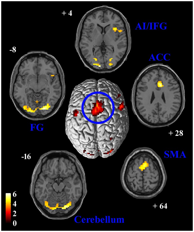
Fig 5. SMA connectivity.
Results of PPI between neural activity in the SMA and the psychological variable of interest (implicit observation of pain expression during temporal processing). The SMA cluster of activation used as seed is displayed at the center, superimposed on a surface rendering of the brain. Areas of significant positive PPI are shown in axial slices of a standard structural T1 weighted brain image. A double statistical threshold was adopted to correct for multiple comparisons, α < 0.05 (3dClustSim): voxel-wise intensity of p < 0.001, and k > 69 voxels. FG = fusiform gyrus, SMA = supplementary motor area, ACC = anterior cingulate cortex, AI = anterior insula, IFG = inferior frontal gyrus.

