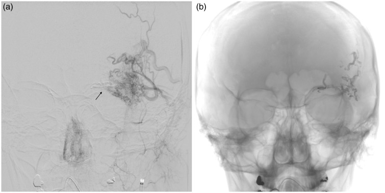
Figure 1.
Cerebral angiogram conducted on the day of embolization. (a) Anteroposterior (AP) view of the left external carotid artery showing supply to the supraorbital arteriovenous malformation from deep and superficial temporal arteries. Arrow shows glue from ophthalmic artery embolization. (b) AP view post-embolization showing glue cast in the arterial pedicles.

