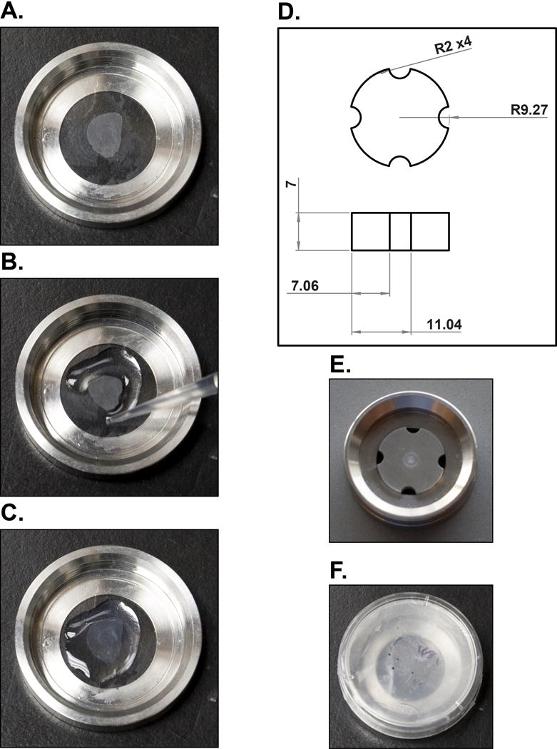
Figure 1.

Brain slice mounting for super-resolution imaging. (A) A brain slice is floated, mounted on circular glass coverslips and then allowed to completely dry. (B, C) Liquid 2% agarose is pipetted along the edges of the brain slice and then across the entire surface to form an immobile pad. (D) Schematics of the stainless steel imaging chamber insert with all dimensions in mm and radii with a curvature measurement of 2 mm. (E) The stainless steel insert is placed in the imaging chamber along with imaging buffer and (F) sealed with a parafilm-lined lid of a 35 mm culture plate for imaging.
