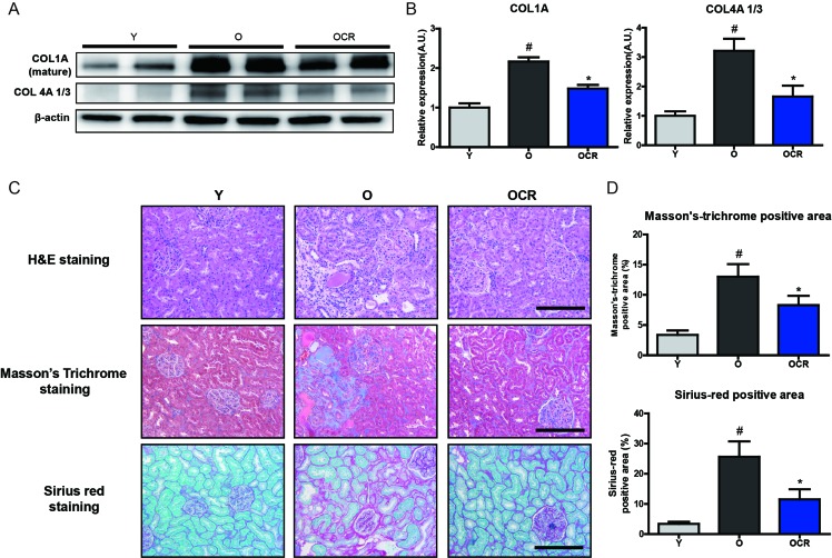
Figure 5. Changes of MMP2 substrate extracellular matrix proteins and fibrosis during aging and CR.
(A) Accumulation of type I and IV collagens during aging in kidney is detected by Western blotting. β-actin was used as loading control. (B) The blots in figure 5A were quantified by densitometry (n = 8). #P < 0.05 vs young. *P < 0.05 vs old. (C) ECM accumulation in young, aged, and CR kidney is visualized by staining with Masson’s Trichrome (MT) and Sirius red staining. Representative pictures of staining results are presented. MT staining and Sirius red staining positive areas were quantified. Both results show that aging increased ECM accumulation and CR attenuates these accumulation. Scale bar = 50 μm. (D) Quantification of Sirius red and MT stained fibrosis in kidneys. #P < 0.05 vs young. *P < 0.05 vs old.

