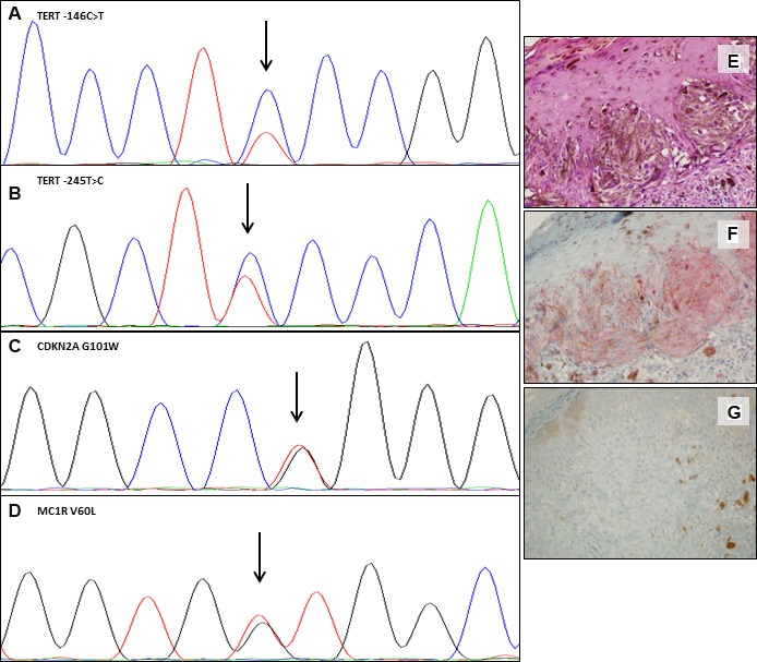
Figure 3. Somatic and germline mutations and variants in one representative case.

Electropherograms showing the TERT promoter –146C>T somatic mutation (A) and the –245T>C polymorphism (B); CDKN2A p.G101W germline mutation (C) and of MC1R p.V60L germline variant (D). Hematoxylin and eosin (E), IHC positive staining for BRAF V600E (F) and IHC showing loss of expression of p16 protein (G). (Magnification ×40). The variant sequence is indicated by an arrow.
