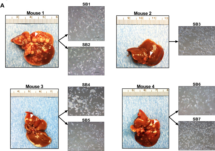
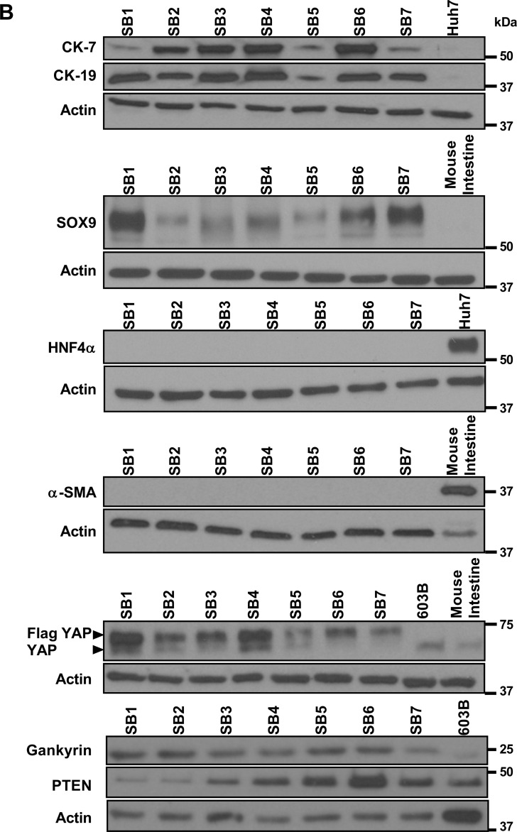
Figure 1. Murine cells derived from YAP-associated tumors have phenotypic features of human CCA.
(A) Liver appearances of mice 10 weeks after having undergone biliary transduction of SB+/–Akt+/–YAP coupled with subsequent systemic IL-33 administration (1 µg i.p. for 3 days) (left panels). Representative phase contrast microscopy images of murine cells derived from distinct tumor nodules (white arrow denotes representative nodule) (right panels). Original magnification 20×. Scale bars: 50 µm. (B) Whole cell lysates were prepared from SB1-7, Huh7, and mouse immortalized, nonmalignant cholangiocytes (603B). Lysates were also prepared from mouse intestine tissue. Lysates were subjected to immunoblot analysis of CK-7, CK-19, SOX9, HNF4α, α-SMA, FLAG-YAP, YAP, gankyrin, and PTEN. β-actin was used as a loading control.


