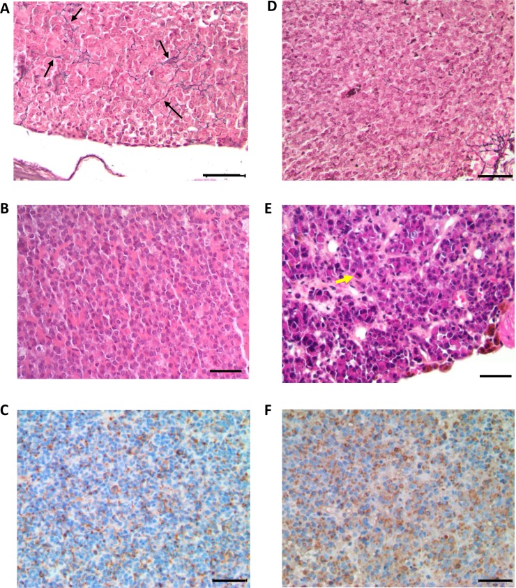
Figure 4. Histological analysis of anterior pituitaries from 24 month-old Prlr+/+ and Prlr–/– mice.
Top row shows Gordon–Sweet silver staining in pituitaries sections of Prlr+/+ (A) and Prlr–/– (D) mice. Black arrows indicate reticulin fibers. Middle row shows hematoxylin and eosin-stained pituitary sections of Prlr+/+ (B) and Prlr–/– (E) mice. The wild-type pituitary has a normal architecture and mixture of cells (B). The Prlr–/– genotype exhibits peliosis (extravasated erythrocytes not contained in capillaries) and lactotrophs with very large Golgi regions and scattered, large, hyperchromatic, atypical nuclei. Yellow arrow indicates mitosis (E). Bottom row: PRL immunohistochemistry. The wild-type pituitary (C) contains a few mature active lactotroph cells with PRL immunostaining. Almost all the cells in the Prlr–/– pituitary (F) are active lactotrophs with PRL immunoreactivity. Bar scale = 50 µm.

