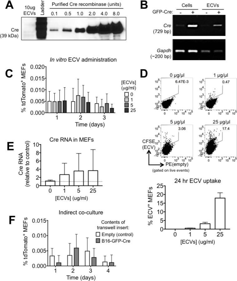
Figure 2. The rapid transfer of Cre from B16 melanoma cells to MEF is not mediated by ECVs.
(A) Analysis of Cre protein in 10 μg of B16-GFP-Cre ECVs by western blotting. (B) Analysis of Cre RNA in B16-GFP-Cre ECVs by PCR. (C) Quantification of the frequency of tdTomato+ MEF after treatment with B16-GFP-Cre ECVs for up to three days (n = 4 independent experiments). Data is represented as mean ± SEM. (D) Representative FACS plots and quantification showing the frequency of MEF that uptake CFSE-labeled ECVs in 24 hrs in vitro (n = 4 independent experiments). Data is represented as mean ± SEM. (E) Analysis of Cre RNA in MEF that were treated with B16-GFP-Cre ECVs for 24 hrs by qPCR. Data were normalized against Hprt (n = 3 independent experiments). Data is represented as mean ± SEM. (F) Quantification of tdTomato expression in reporter MEF that were cultured alone (control) or indirectly with B16-GFP-Cre cells (separated by a membrane with 0.4μm pores) for up to 4 days (n = 4 independent experiments). Data is represented as mean ± SEM. See also Supplementary Figure 1.

