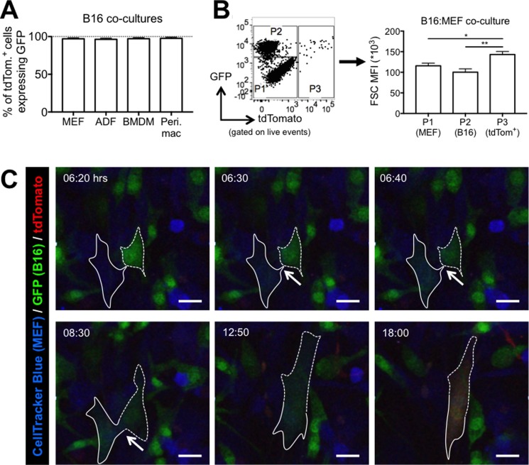
Figure 3. Cell-cell fusion mediates the rapid transfer of bioactive Cre from B16 melanoma cells to non-cancer cells in vitro.
(A) Quantification of GFP expression in tdTomato+ cells from 48 hour co-cultures of B16-GFP-Cre cells with various reporter cells (n = 3–4 independent experiments). Data is represented as mean ± SEM. (B) Quantification of FSC MFI of three populations of cells from a 24 hour B16:MEF co-culture: P1 = GFP–, tdTomato- (MEF); P2 = GFP+ (B16 cells); P3 = tdTomato+ (MEF that received bioactive Cre) (n = 7 independent experiments). Data is represented as mean ± SEM. See also Supplementary Figure 2. (C) Stills from confocal imaging movie (Video 1) of B16:MEF co-culture showing a CellTracker Blue-labeled reporter MEF (outlined in solid white line) turn green and then red after fusing with a B16-GFP-Cre cell (outlined in dashed white line). Arrows indicate the area of contact between the MEF and B16 cell that ultimately fuse and start expressing tdTomato at 18:00 hrs. See also Supplementary Videos 1–5.

