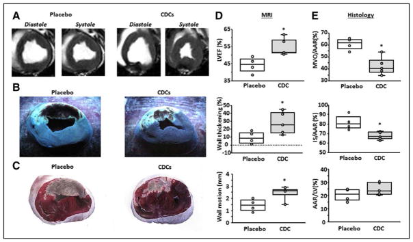
Figure 1. Validation of cellular postconditioning in pigs.
A, MR short-axis images from a placebo and CDC-treated pig. Transverse cardiac slices stained with Thioflavin T and Gentian Violet (B), and triphenyl tetrazolium chloride (TTC) (C) in the same representative sample. B, The area of MVO appears nonfluorescent under UV light, whereas the area-at-risk (AAR) is unstained with Gentian Violet. C, Viable myocardium appears red and scar appears white/yellow. LVEF (D, top), infarct wall thickening (D, middle), and infarct wall motion (D, lower) are improved following CDC treatment. MVO/AAR (E, top), and infarct size (IS)/AAR (E, middle) are decreased following CDC treatment, whereas AAR is not different between groups (E, lower). Graphs depict mean±SEM. Statistical significance was determined by using the Student t test. *P<0.05. CDC indicates cardiosphere-derived cell; LVEF, left ventricular ejection fraction; MVO, microvascular occlusion; and SEM, standard error of the mean.

