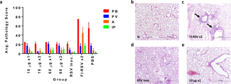
Figure 4. Cotton rat lung pulmonary histopathology after RSV/A challenge.

The lungs of Cotton rats in groups A-G were harvested 5 days after 1×10⁵ RSV/A challenge. Lungs were formalin fixed, sectioned and H&E stained. Slides were scored on a 0-4 severity scale in a blinded manner. Scores were converted onto a 0-100% histopathology scale (0=no; 25=minimal, 50=mild; 75=moderate; 100=maximal inflammation). The parameters evaluated were peribronchiolitis (PB), perivasculitis (PV), alveolitis (A) and interstitial pneumonia (IP). Bars represent mean pathology scores + SEM (a). Representative lung H&E images non-treated animal (b), and 5 days post challenge from FI-RSV vaccinated animal (c), day 0 RSV/A infected animals (d) and from pGX2303 immunized animal from (e). Magnification x40.
