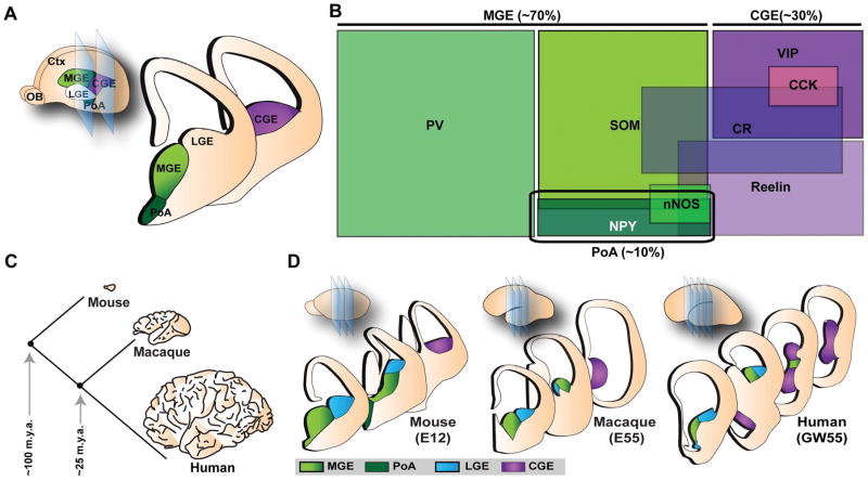
Figure 2. Developmental origins and diversity of cortical interneurons.
(A) Cortical interneurons in mouse are derived from progenitor cells located in the proliferative zones of the ventral telencephalon, specifically in the MGE (medial ganglionic eminence), CGE (caudal ganglionic eminence) and PoA (preoptic area). LGE, lateral ganglionic eminence; Ctx, cortex; OB, olfactory bulb. (B) The major source of interneurons is the MGE, generating ~70% of cortical interneurons comprised of two non-overlapping populations expressing PV (parvalbumin) and SOM (somatostatin). ~30% of cortical interneurons are a heterogenous group of cells generated by the CGE, all of which express 5HTR (serotonin receptor)-3A as well as either VIP (vasointestinal peptide) or reelin. In addition, CGE is the main source of CR (calretinin) and CCK (cholecystokinin)-expressing cells. The PoA generates ~10% of interneurons a fraction of which express NPY (neuropeptide Y), nNOS (neuronal nitric oxide synthase), and SOM. (C) Evolution of the mammalian brain from lower mammals such as mouse to primates such as macaque monkey and human. Mouse and macaque monkey diverged from human ~100 and ~25 m.y.a. (million years ago), respectively. Modified from Rakic, P. 2009.150 (D) Developmental origins of cortical interneurons across mammalian species. Inspired from Molnar, Z. et al., 2013.151 E, embryonic day; GW, gestation week.

