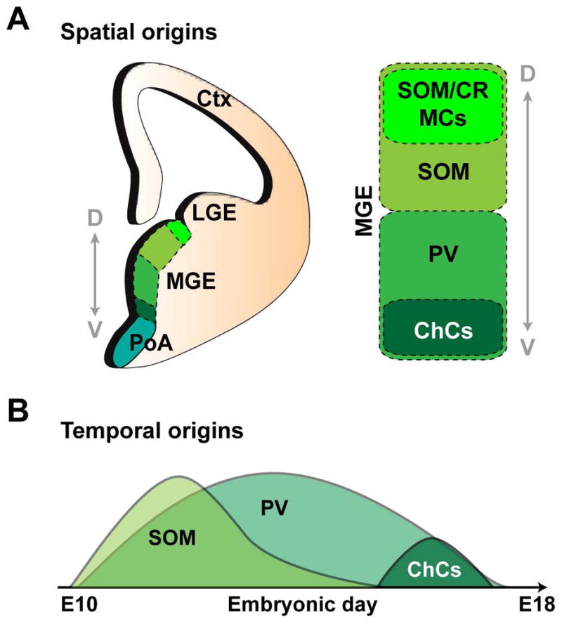
Figure 3. Spatial and temporal origins of MGE-derived interneurons.

(A) Spatial bias of progenitors to generate SOM (somatostatin)- and PV (parvalbumin)- expressing cells in the dorsal and ventral regions of the MGE (medial ganglionic eminence), respectively. SOM/CR (calretinin)- expressing cells have been suggested to originate in the dorsal-most tip of the MGE, where as ChCs (chandelier cells) are enriched in ventral MGE. LGE, lateral ganglionic eminence; PoA, preoptic area; Ctx, cortex. (B) Temporal bias in MGE fate-specification. Whereas the production of SOM+ cells peaks early and subsequently declines, PV+ cells are continuously generated throughout embryonic neurogenesis. ChCs are preferentially born at the very late embryonic stage. Adapted from Bandler, R.C. et al., 2017.152
