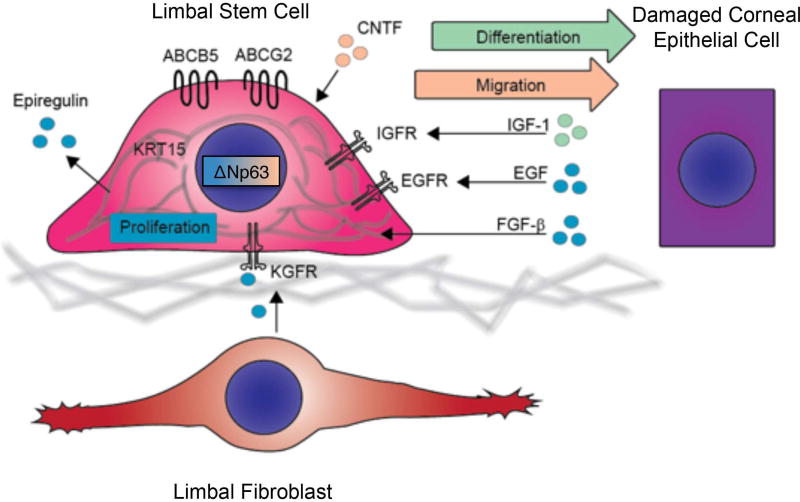
Figure 5. Factors affecting corneal epithelial wound healing.
Normal corneal wound healing is dependent on proliferation (blue), migration (orange) and differentiation (green) of corneal progenitors. Attenuated expression of LSC markers, including ABCB5, ABCG2, ΔNp63α or K15, is associated with abnormal corneal wound healing, which may result in increased corneal fragility, ulceration and clouding. Limbal epithelial cell proliferation is supported by expression of ΔNp63α and epiregulin in limbal basal epithelial cells, by keratinocyte growth factor (KGF) secreted from limbal fibroblasts, and by epidermal growth factor (EGF) and fibroblast growth factor-β (FGF-β) produced by damaged corneal epithelium. Migration is promoted by expression of ΔNp63α and ciliary neurotrophic factor (CNTF). Differentiation is induced by insulin-like growth factor-I (IGF-I), rapidly produced by injured corneal epithelium upon injury. KGFR, KGF receptor; EGFR, EGF receptor; IGFR, IGF receptor.

