
Figure 4.
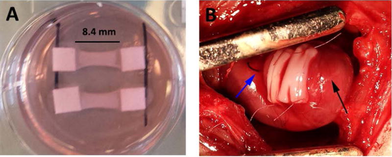
Bi-layer patches at implantation. A) Patches prior to removing from spacers after two weeks of culture. B) Two patches sutured onto the left ventricular epicardial surface after myocardial infarction. Blue arrow indicates ligation suture, black arrow indicates apex of heart.