Fig. 3.

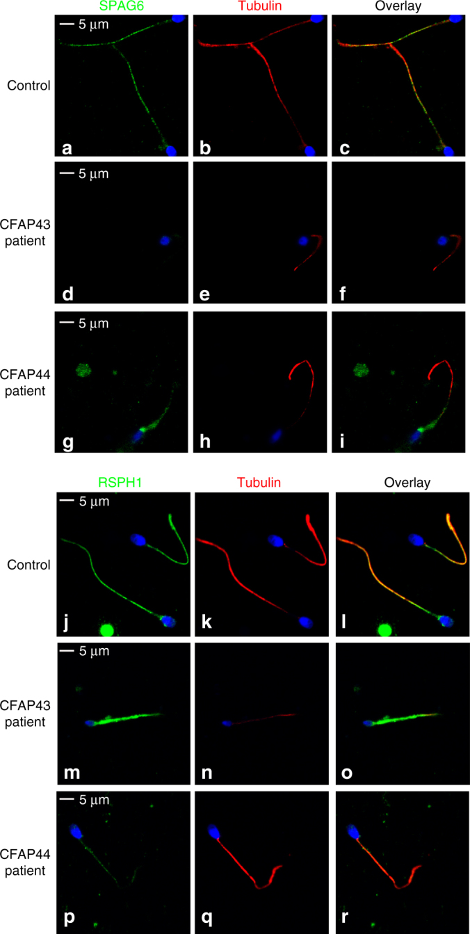
Immunofluorescence staining in CFAP43 and CFAP44 patients reveals an abnormal axonemal organization. a–c sperm cells from a fertile control stained with anti SPAG6 (green), a protein located in the CPC, and anti-acetylated tubulin (red) antibodies. DNA was counterstained with Hoechst 33342. c Corresponds to a and b overlay and shows that in control sperm, SPAG6 and tubulin staining superimpose. Scale bars = 5 µm. d–f SPAG6 staining is absent in sperm from the patient P43-5 homozygous for the c.2658C>T variant in CFAP43. d–i Similar IF experiments performed with sperm cells from the patient P44-2 homozygous for the c.3175C>T variant in CFAP44. Scale bar = 5 µm. Contrary to the control, the SPAG6 immunostaining (green) is abnormal with a diffuse pattern concentrated in the midpiece of the spermatozoa and is not detectable in the principle piece. j–l Sperm cells from a fertile control stained with anti RSPH1 (green), a protein of the radial spoke’s head, and anti-acetylated tubulin (red) antibodies. DNA was counterstained with Hoechst 33342. l corresponds to j and k overlay and shows that RSPH1 and tubulin staining superimpose in control sperm. Scale bar = 5 µm. m–o In sperm from the patient P43-5, RSPH1 staining (green) is significantly different from control (m) with a marked diffuse staining. p–r In sperm from the patient P44-2 the intensity of the RSPH1 staining is strongly reduced
