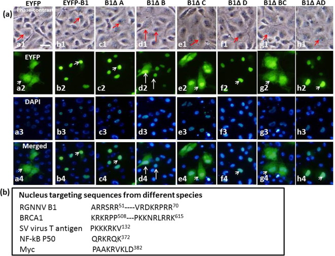
Figure 5.
Fluorescence microscopy and DAPI staining confirm the role of domains B and C in nuclear targeting of B1 in GF-1 cells. (a) Phase-contrast fluorescence images showing the distribution of EYFP, EYFP-B1 and different B1 domain deletion mutants in transfected GF–1 cells at 48 hpt. Panels a1–a4 show EYFP distributed in the cytoplasm; panels b1–b4 show EYFP-B1 distributed in the nucleus; panels c1–c4 show EYFP-B1ΔA distributed in the nucleus; panels d1–d4 show EYFP–B1ΔB distributed in the cytoplasm (major) and nucleus (minor; indicated by arrows); panels e1–e4 show EYFP–B1ΔC distributed in the cytoplasm (indicated by arrows); panels f1–f4 show EYFP–B1ΔD distributed in the nucleus; panels g1–g4 show EYFP–B1ΔBC distributed in the cytoplasm; panels h1–h4 show EYFP–B1ΔAD distributed in the nucleus. The cells were also traced with green fluorescence and DAPI nuclear staining and the final images were merged to confirm localization. Scale bar = 10 μM. (b) Comparison of different nuclear targeting signal sequences.

