Fig. 3.
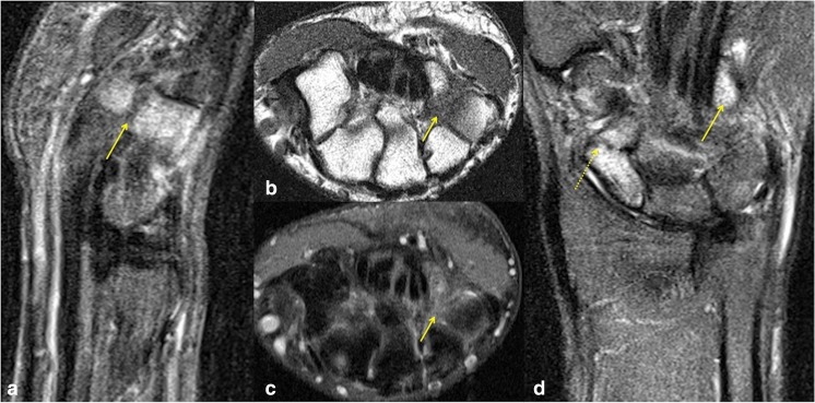
A 21-year-old man with fracture of the left scaphoid and concomitant hook of hamate fracture. a Sagittal STIR shows significant marrow oedema involving the hook and body of the hamate, which is easily discernible compared with the low signal observed in the other imaged bones. A low signal fracture line is seen across the base of the hook of hamate (arrow). b Hook of hamate fracture on axial T1 and c corresponding axial STIR sequences, both demonstrating obvious marrow signal abnormality, although the displaced fracture line is more conspicuous on T1. d Coronal STIR sequences easily demonstrate marked oedema with angulated fracture of the distal scaphoid pole (dashed arrow), but also draw attention to marked marrow oedema of the hook of hamate (arrow). As is generally the rule with MRI of most fractures, bony abnormality was most notable on the fluid-sensitive sequences due to marrow oedema, although the fracture line was most conspicuous on T1

