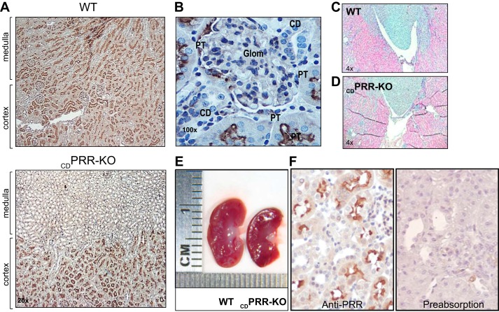
Fig. 1.
A: kidney sections (3 µm) from a wild-type (WT) mouse (top) showed positive prorenin receptor (PRR) immunoreactivity in glomeruli, proximal tubules, and collecting ducts (CDs) in renal cortex and medulla; however, in the kidney from a deletion of PRR in the CD (CDPRR-KO) mouse (bottom), PRR immunoreactivity was mostly detected in cortical tubules but absent in renal medulla, primarily containing collecting ducts (×20 magnification). B: high magnification of PRR immunostaining (3,3prime diaminobenzidine tetrahydrochloride; DAB; brown chromogen, ×100) in a kidney section (3 µm) from a CDPRR-KO mouse. PRR was detected in glomeruli and proximal tubules (PT) but not in the CDs of CDPRR-KO mouse kidney. Kidney sections (3 µm, ×4 magnification) hematoxylin-stained from a wild-type (WT) (C) and knockout (KO) mice (D). E: gross morphology analysis in kidneys from a WT mouse and CDPRR-KO showing kidney size and internal structure. No evidence of significant medullary hypoplasia was observed. F: preabsorption of primary PRR antibody for 72 h using preimmune peptide 30-fold excess denotes the specificity of the antibody. Glom, glomeruli.

