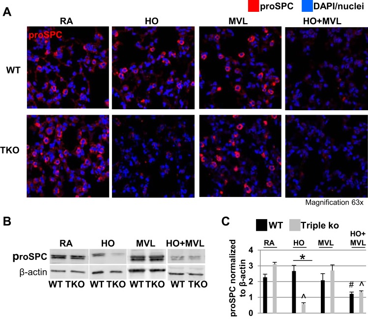
Fig. 3.
HO exposure, but not MVL, decreased proSPC protein levels in TREK-1/TREK-2/TRAAK-deficient lungs. A: representative confocal immunofluorescence microscopy images showing that 95% HO exposure for 72 h decreased proSPC protein levels in triple KO but not WT control lungs. MVL alone had no effect on either mouse type. HO + MVL decreased proSPC protein levels in both WT control and triple KO mice. ProSPC protein is shown in red, and nuclei were counterstained in blue with DAPI (n = 5 mice per treatment group). B: representative Western blots using whole lung homogenates showing the effects of the above conditions on proSPC protein expression. For a specific treatment condition, tissue lysates from WT control and triple KO lungs were run on the same gel to allow for comparison of protein expression levels between the 2 groups. C: normalized densitometry analysis of Western blot experiments (n = 4–5 mice per group; *comparison of WT control and triple KO mice within 1 treatment group; #compared with untreated WT control mice; ^compared with untreated triple KO mice).

