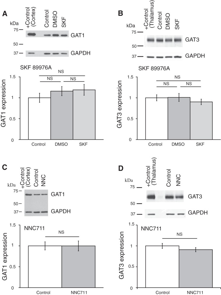
Fig. 7.
GAT1 inhibitors SKF-89976A and NNC-711 did not affect the expression of GAT1 and GAT3 in the hypothalamus. Brain slices were incubated for 5–7 h in ACSF containing SKF-89976A (SKF; 100 µM) dissolved in DMSO (0.1% final) or NNC-711 (NNC; 5 µM) dissolved in water. The brain tissue was then processed for Western blot analysis. A and B: effect of SKF; n = 6 triplicates: control, DMSO (vehicle), and SKF. C and D: effect of NNC-711; n = 8 duplicates: control and NNC-711. Bands above graphs show GAT1 (A and C) or GAT3 (B and D) expression and loading control (GAPDH). +Control, positive controls (cerebral cortex for GAT1 and thalamus for GAT3). The optical density of bands was quantified with ImageJ for each gel, normalized to the loading control, and shown as a ratio to control on bar graphs (means ± SE; one-way ANOVA, Tukey HSD post hoc test or t-test; NS, nonsignificant changes). GAT1 inhibitors did not change the expression of either GAT [A: F2,15 = 0.73 (Fcrit = 3.68), P = 0.50; B: F2,15 = 0.56 (Fcrit = 3.68), P = 0.59; C: F1,14 = 8E-05 (Fcrit = 4.60), P = 0.99; D: F1,14 = 1.43 (Fcrit = 4.60), P = 0.25]. DMSO did not significantly affect the expression of the GAT proteins.

