
Figure 8.
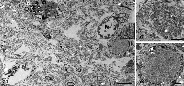
Human reactive astrocytes in Alzheimer's brains phagocytose dystrophic neurites. TEM of post mortem human Braak V–VI tissue showing a reactive astrocyte containing dystrophic neurites (a1). Small bundles of filaments are observed (arrows). Higher magnification views of the squared areas in c showing dystrophic neurites filled with autophagic vesicles (a2) or containing paired‐helical filaments (a3). N, astrocyte nucleus. Scale bars: a1, 5 µm; a2 and a3, 1 µm