
Figure 9.
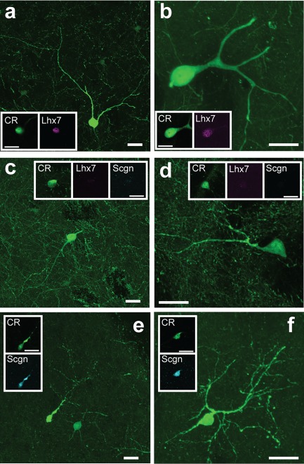
The selective expression of Scgn or Lhx7 identifies morphologically distinct “types” of CR‐expressing interneurons in mouse striatum. (a–f) Confocal micrographs of the mouse dorsal striatum showing the different morphological characteristics of CR‐expressing interneurons. (a, b) CR+/Lhx7+ (“Type 1”) interneurons had large somata with 3 or more relatively thick primary dendrites. (c, d) CR+/Scgn−/Lhx7− (“Type 2”) interneurons had medium‐sized somata and often had bipolar dendrites. (e, f) CR+/Scgn+/Lhx7− (“Type 3”) interneurons had small somata and tortuous dendrites. For (a–f), large panels show maximum intensity projections taken across multiple optical planes (z‐stacks) containing the extent of the labeled neuron's somatodendritic structure, while the boxed insets are single‐plane images of the somata of the same neurons. All scale bars are 20 µm