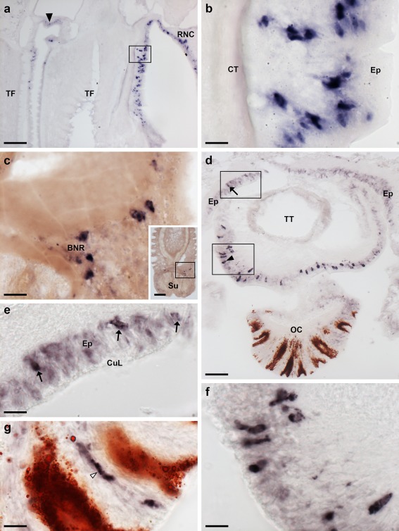
Figure 4.

Localization of ArPPLNP2 mRNA in tube feet and the arm tip region of A. rubens using in situ hybridization. (a) Longitudinal section of a tube foot showing stained cells at the junction with an adjacent tube foot (arrow) and in the sub‐epithelial layer near to the junction with the adjacent radial nerve cord (rectangle), which is shown at higher magnification in (b). (c) Stained cells located near to the tube foot basal nerve ring. The inset shows the location of the stained cells (rectangle) in a lower magnification image of the tube foot. (d) Transverse cryostat section of an arm tip showing the pigmented optic cushion and terminal tentacle. Stained cells can be seen in the terminal tentacle external epithelium (rectangle with arrowhead) and in the body wall epithelium (rectangle with arrow). The boxed areas in (d) are shown at higher magnification in (e) and (f). (g) High magnification image of the photoreceptor cell layer of an optic cushion showing a stained cell (white arrowhead) between pigmented cells. BNR, basal nerve ring; CT, collagenous tissue; CuL, cuticle layer; Ep, epithelium; OC, optic cushion; RNC, radial nerve cord; Su, sucker; TF, tube foot; TT, terminal tentacle. Scale bars: 100 μm in (a), (c) inset; 10 μm in (b), (c), (e), (f), (g); 50 μm in (d)
