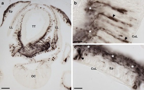
Figure 8.

Localization of ArPPLN2h immunoreactivity in the terminal tentacle and associated structures in the arm tip of A. rubens. (a) Immunostaining in a transverse section at the base of the terminal tentacle. Stained cells and processes can be seen here in the body wall epithelium, the terminal tentacle and associated lateral lappets and the optic cushion. (b) A high magnification image of arm tip epithelium showing immunostained bipolar cells in the epithelium (arrowhead) and in a dense meshwork of fibers in the underlying basiepithelial plexus (asterisks). (c) High magnification image of the terminal tentacle showing immunostained bipolar cells in the epithelium (arrowheads) with stained processes projecting into a dense meshwork of fibers in the underlying basiepithelial plexus (asterisks). CuL, cuticle layer; Ep, epithelium; LL, lateral lappet; TT, terminal tentacle; OC, optic cushion. Scale bars: 100 μm in (a); 10 μm in (b, c)
