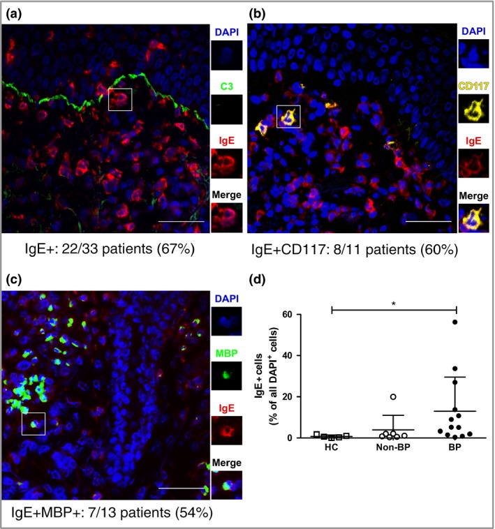
Figure 2.

IgE is found in the perilesional skin of patients with BP, mostly bound to mast cells and eosinophils. Direct immunofluorescence of BP perilesional skin for (a) IgE, (b) mast cells (CD117+ ) and (c) eosinophils (MBP+). The number of patients tested and found positive for each case is indicated. Scale bars = 50 μm. (d) IgE+ cells were calculated for healthy controls (HCs) (n = 5), non‐BP disease controls (n = 7) and patients with BP (n = 13). Non‐BP disease controls include linear IgA disease, vasculitis, epidermolysis bullosa acquisita, pemphigus and dermatitis herpetiformis. Positive cells are presented as a percentage of all dermal cells identified by 4′,6‐diamidino‐2‐phenylindole (DAPI) staining. The data represented in the graph are mean ± SD. *P < 0·05. MBP, major basic protein.
